
For anatomy students, the hardest part of learning phalanges is rarely the number itself. The challenge is connecting the number to the terminology: proximal versus distal, PIP versus DIP, phalanx versus metacarpal, and thumb versus the other fingers.
Once those relationships are organized into a pattern, the topic becomes much easier.
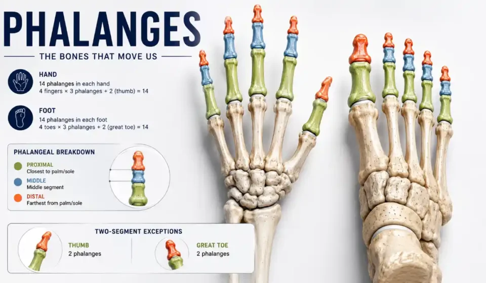
In standard anatomy, each hand has 14 digit bones, and each foot has 14. Four fingers and four lesser toes have three segments each, while the thumb and great toe have two.
This guide is organized around the relationships most useful for studying, recalling, and applying the anatomy rather than simply presenting a list of definitions.
Start With the Number Pattern
The basic formula is:
4 × 3 + 2 = 14
For one hand:
- Index finger = 3
- Middle finger = 3
- Ring finger = 3
- Little finger = 3
- Thumb = 2
That gives:
12 + 2 = 14
The same calculation applies to the foot:
- Second toe = 3
- Third toe = 3
- Fourth toe = 3
- Fifth toe = 3
- Great toe = 2
Again:
12 + 2 = 14
The hands and feet therefore contain 56 digit bones altogether.
| Region | Three-segment digits | Two-segment digit | Total |
| One hand | 4 × 3 | Thumb × 2 | 14 |
| Both hands | 8 × 3 | 2 thumbs × 2 | 28 |
| One foot | 4 × 3 | Great toe × 2 | 14 |
| Both feet | 8 × 3 | 2 great toes × 2 | 28 |
Read More: Phalanges
This is more useful than memorizing “14” because you can reconstruct the answer if you forget it.
Proximal, Middle, and Distal: Think in Position
The terminology becomes easier if you treat it as a directional system.
Proximal means closer to the hand or foot.
Middle means between the other two segments.
Distal means farther from the hand or foot, toward the digit tip.
A typical finger therefore follows:
Metacarpal → proximal → middle → distal
A typical lesser toe follows:
Metatarsal → proximal → middle → distal
The thumb and great toe use:
Metacarpal/metatarsal → proximal → distal
That last line is the exception students need to remember.
Middle and Intermediate Mean the Same Thing
Some anatomy texts use middle phalanx, while others use intermediate phalanx.
For the standard three-segment digits, these terms refer to the same bone.
They should not be treated as two separate structures.
Hand and Foot: The Same Pattern With Different Demands
The hand and foot contain the same number of digit bones, but their functions are very different.
| Feature | Hand | Foot |
| Total | 14 | 14 |
| Two-segment digit | Thumb | Great toe |
| Three-segment digits | Four fingers | Four lesser toes |
| Base articulation | MCP | MTP |
| Primary role | Dexterity and manipulation | Stability and locomotion |
| Larger supporting region | Wrist/palm | Ankle/midfoot/forefoot |
Read More: Phalanges
The hand needs to position the digits around objects. The foot must repeatedly manage body weight, ground contact, balance, and forward movement.
This is why it is better to say that the two regions share a structural pattern, not that they are functionally identical.
Where the Digit Bones Fit
A useful exam sequence is:
Hand: carpals → metacarpals → digit bones
Foot: tarsals → metatarsals → digit bones
The five metacarpals form the skeletal framework of the hand beyond the wrist. The five metatarsals occupy the forefoot and connect distally to the bases of the toes.
This produces two common exam distinctions:
Metacarpal ≠ phalanx
Metatarsal ≠ phalanx
A metacarpal belongs to the hand proper. A metatarsal belongs to the forefoot. The phalanges are the bones of the fingers and toes.
The Joints You Need to Know
Joint terminology is easier when matched directly to the bone relationship.
| Joint | Location | Bones involved |
| MCP | Base of finger | Metacarpal + proximal segment |
| MTP | Base of toe | Metatarsal + proximal segment |
| PIP | Middle of three-segment digit | Proximal + middle |
| DIP | Near digit tip | Middle + distal |
| IP | Thumb/great toe | Proximal + distal |
Read More: Phalanges
The index, middle, ring, and little fingers have PIP and DIP joints. The thumb has only one IP joint because there is no middle segment. The lesser toes follow the three-segment pattern, while the great toe has one IP joint.
A high-yield rule is:
Three bones = two interphalangeal joints. Two bones = one interphalangeal joint.
Base, Shaft, Head, and Tuft
For the proximal and middle bones, the basic structural description is:
Base → shaft → head
The base is closest to the hand or foot. The shaft forms the central portion. The head forms the joint surface at the other end.
The distal bone is different. It has a base and shaft followed by a terminal tuft, which is associated with the soft tissues at the end of the finger or toe. In the finger, the distal bone is closely related to the nail plate and nail bed.
That difference is worth remembering because a simplified diagram showing every digit bone as identical can create a misleading picture.
What Moves the Fingers?
Bones provide the rigid framework, but muscles and tendons generate movement.
Three relationships are particularly useful for students:
FDS → middle phalanx → PIP flexion
FDP → distal phalanx → DIP flexion
Extensor mechanism → finger extension
The flexor digitorum superficialis (FDS) inserts on the middle segment and is a major flexor of the PIP joint.
The flexor digitorum profundus (FDP) extends farther distally and inserts on the distal segment, allowing it to flex the DIP joint.
The extensor mechanism coordinates extension through a more complex arrangement of tendons and expansions.
This is an example of a better study strategy: learn the attachment and action relationship instead of memorizing tendon names in isolation.
Exam Traps Students Commonly Miss
Trap 1: Giving the Thumb Three Bones
The thumb has two.
Trap 2: Giving the Great Toe Three Bones
The great toe has two.
Trap 3: Calling the Middle Segment a Fourth Type
“Middle” and “intermediate” describe the same segment.
Trap 4: Giving the Thumb PIP and DIP Joints
The thumb has one IP joint.
Trap 5: Calling the Metacarpal a Digit Bone
The metacarpal belongs to the hand skeleton immediately proximal to the digits.
Trap 6: Assuming Every Bone Has the Same Shape
Proximal and middle segments have a base, shaft, and head, whereas the distal segment has a specialized terminal tuft.
Trap 7: Saying the Foot Has Different Numbers
Each foot also has 14.
A Better Way to Solve Anatomy Questions
Suppose an exam asks:
Which tendon inserts on the bone that flexes the DIP joint?
Do not try to recall the entire hand anatomy chapter.
Start with the joint:
DIP
Then identify the bone that moves at that joint:
Distal
Then identify the major flexor:
FDP
The answer becomes:
Flexor digitorum profundus.
The same reasoning works in reverse.
If a question mentions the middle phalanx, think:
PIP → FDS
If it mentions the distal phalanx and fingertip flexion, think:
DIP → FDP
This is more robust than memorization because it links anatomy to function.
Why the Thumb Needs Separate Attention
The thumb is not simply a shorter version of another finger.
It has two digit bones and a specialized first carpometacarpal joint that permits movements important for opposition and grasping. The first MCP joint also has sesamoid bones that contribute to its mechanical environment.
For exam purposes, however, the essential distinction remains:
Thumb: proximal + distal
That missing middle segment changes the interphalangeal joint pattern as well.
Why the Great Toe Needs Separate Attention
The great toe follows the same two-bone pattern as the thumb, but its mechanical role is different.
The first metatarsal connects to the proximal segment of the hallux at the first MTP joint. During gait, the great toe participates in the transition from weight acceptance to propulsion. The foot as a whole functions through coordinated interactions among bones, joints, muscles, tendons, and soft tissues.
This is why an anatomy answer should not reduce the great toe to simply “the foot’s thumb.”
The two structures share a two-bone pattern, but their functional environments are very different.
Read More: Phalanges
Why This Anatomy Matters Clinically
Understanding the normal arrangement helps explain injuries.
Phalanx fractures can be described according to whether they affect the base, shaft, or head/condylar region and whether alignment or joint surfaces are involved.
The surrounding tendons and ligaments also matter. A fracture may be mechanically important not only because of the broken bone itself but because altered alignment can interfere with joint movement or tendon function.
Mallet finger is a useful example. It involves disruption of the extensor mechanism at the distal end of the finger and may occur with an avulsion fracture.
The clinical lesson is straightforward:
Bone anatomy explains the structure; tendon and joint anatomy help explain the movement.
The Five-Line Study Sheet
If you had to reduce the topic to five lines before an exam, use this:
14 per hand.
14 per foot.
Four ordinary digits = 3 each.
Thumb/great toe = 2 each.
Proximal → middle → distal; MCP/MTP → PIP → DIP.
That compact framework lets you reconstruct most of the basic anatomy without memorizing dozens of unrelated statements.
Frequently Asked Questions
How many bones are in each finger?
The index, middle, ring, and little fingers each contain three digit bones. The thumb contains two.
How many digit bones are in one hand?
There are 14: twelve across the four three-segment fingers and two in the thumb.
Is the intermediate phalanx different from the middle phalanx?
No. They are alternative terms for the central bone in a three-segment digit.
Which joint is between the proximal and middle segments?
The PIP joint, or proximal interphalangeal joint.
Which joint is between the middle and distal segments?
The DIP joint, or distal interphalangeal joint.
Why does the thumb have one IP joint?
Because it has only proximal and distal segments, which meet directly at the interphalangeal joint.
Which tendon inserts on the middle phalanx?
The flexor digitorum superficialis inserts on the volar base of the middle phalanx and contributes to PIP flexion.
Which tendon inserts on the distal phalanx?
The flexor digitorum profundus inserts on the distal phalanx and flexes the DIP joint.
Are hand and foot digit bones counted the same way?
Yes. Each hand and each foot normally has 14, although their functional roles differ.
What is the easiest way to remember the number?
Use 4 × 3 + 2 = 14. Four ordinary digits have three bones each, and the exceptional digit has two.